What Is the Basic Pathological Change With Macular Degeneration
Degenerationdysregulation of the retinal pigment epithelium RPE a supportive monolayer of cells underlying the photoreceptors is commonly seen in patients with AMD. Degeneration of the retinal cells in the fovea centralis d.

Macular Degeneration Physiopedia
In a case of acute otitis media what would a purulent discharge in the external canal of the ear and some pain.

. Age-related macular degeneration is an eye disease that may get worse over time. Macular changes in dry AMD are characterized by subretinal drusen deposits atrophy of the retinal pigment epithelium RPE pigment epithelial detachments and subretinal pigment epithelial clumping. Macular Degeneration Degenerative changes in the RETINA usually of older adults which results in a loss of vision in the center of the visual field the MACULA LUTEA because of damage to the retina.
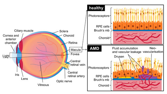
Overall the pathological mechanisms proposed in AMD can be divided into 4 categories of inflammation oxidative stress abnormal ECM production formation of CNVs and neovascularisation Zarbin 2004. Excess metabolic byproducts form and accumulate in the retinal pigment epithelium leading to the formation of drusen. Sorsbys Fundus Dystrophy SFD is a rare autosomal dominant maculopathy.
Agerelated macular degeneration AMD is a progressive degenerative disease that is the leading cause of vision loss in the elderly population. Age-related macular degeneration AMD was first described in the medical literature in 1875 as symmetrical central choroidoretinal disease occurring in senile persons Hutchison and Tay 1875. The basic change in macular degeneration is.
Age-Related Macular Degeneration Diagnosis. Learn more about the symptoms tests. While it does not result in complete blindness loss.
The heredity of this disorder is autosomal dominant with reduced penetrance and variable expressivity. With the advent of aging society of China fundus diseases related to pathological neovascularization including age-related macular degeneration AMD diabetic macular edema DME and pathological myopia PM have become an increasingly serious medical and health problems. Movement of vitreous humor between the retina and the choroid c.
Over time however some people experience a gradual worsening of vision that may affect one or both eyes. Pathologic changes occur primarily in the retinal pigment epithelium Bruchs membrane and choriocapillaries in the macular region that result from the hardening and obstruction of retinal arteries. Although the etiology and pathogenesis of AMD remain largely unclear a complex interaction of genetic and environmental factors is thought to exist.
Pathological changes in the choroidal vasculature underneath the macula lead to NVAMD. Degeneration of the retinal cells in the fovea centralis. There is an absence of neovascularization 3.
AMD pathology is characterized by degeneration involving the retinal photoreceptors retinal pigment epithelium and Bruchs membrane as well as in some cases alterations in choroidal capillaries. Macular degeneration rarely causes total blindness because only the center of vision is affected. He or she can see if there are changes in the retina and.
Early on there are often no symptoms. It is marked by central vision loss with peripheral vision relatively spared. All of the above.
Dry age-related Macular Degeneration. According to the most recent report on the causes of visual impairment by the World Health Organization in 2002 AMD is among the most common causes of blindness. However injury to the macula in the center of the retina can impair the ability to see straight ahead clearly and sometimes make it difficult.
About 5 of people with pathological myopia will start to develop abnormal new blood vessels underneath the macula and if those blood vessels begin to leak a person will be diagnosed with myopic choroidal neovascularizationmyopic macular degeneration. An eye disease that progressively destroys the macula the central portion of the retina impairing central vision. What is the basic pathologic change with macular degeneration.
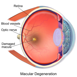
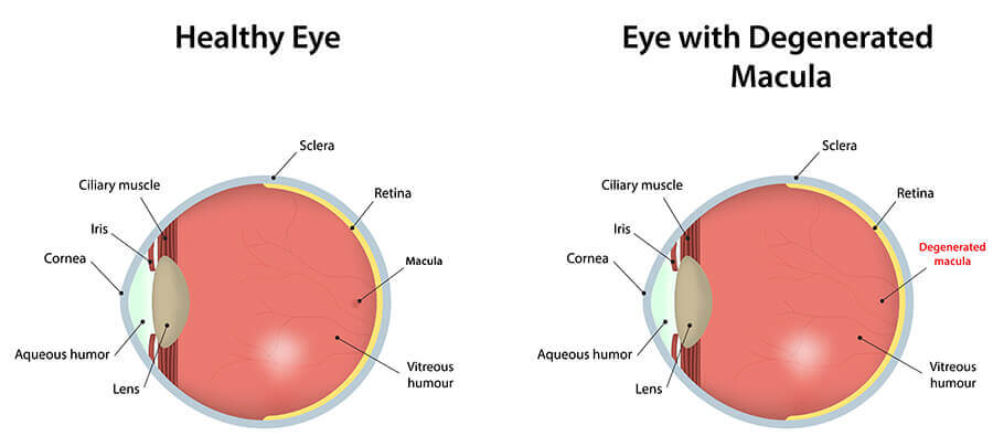
Macular degeneration also known as age-related macular degeneration AMD or ARMD is a medical condition which may result in blurred or no vision in the center of the visual field. Damage to the optic nerve and meninges e. There are two basic types of macular degeneration.
Age-related macular degeneration AMD is visual impairment due to changes in the macula the area responsible for high-acuity vision. Human macular choriocapillaries is estimated to be only 11 as opposed to 94 for retinal capillaries 12. While age-related macular degeneration and myopic macular.
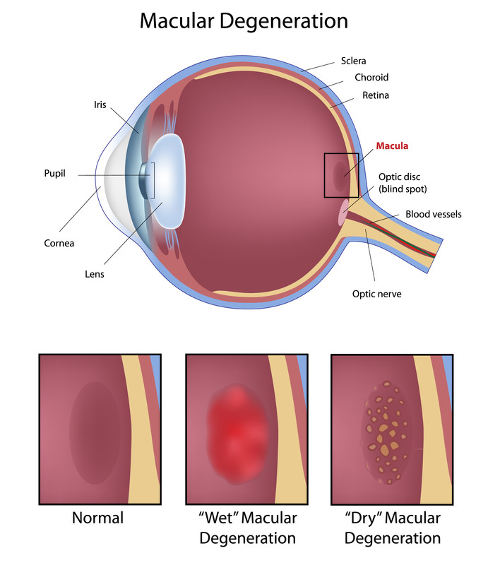
Approximately 10-15 of the cases of macular degeneration are the wet exudative type. Age-related macular degeneration AMD is a leading cause of irreversible blindness in the world. Which of the following is an example of conduction deafness.
Dry AMD affects 85 to 90 percent of everyone with AMD 4. Age a progressive deterioration of the portion of the retina called the macula lutea resulting in loss of central vision. Adhesions reducing the movement of the ossicles.
What is the basic pathological change with macular degeneration. Increased amount of aqueous humor in the eye b. Any changes in the macula or any degeneration or dying of cells of the macula results in vision changes but not in memory loss.
Recent research on the genetic and molecular underpinnings of AMD brings to light several basic molecular pathways and. This grid helps you notice any blurry distorted or blank spots in your field of vision. Your ophthalmologist will also look inside your eye through a special lens.
As effective drugs of the treatment conbercept and ranibizumab. It happens when a part of the retina called the macula is damaged. With AMD the patient loses their central vision but the patients peripheral side vision will still be normal.
Some contradictions exist regarding the nature of the primary defect in this entity. Electrooculographic and angiographic investigations lend support to the belief that the basic pathological changes are located in the retinal pigment epithelium. These unique features may render the microvessels in the choroid more prone to undergo structural changes if the system is stressed.
During an eye exam your ophthalmologist may ask you to look at an Amsler grid. AMD pathology is characterized by degeneration invol. Its the leading cause of severe vision loss in people over age 60.
Sorsby first described it in 1949 when he identified five families whose members developed central visual loss before their forties and whose fundal appearances resemble that of patients suffering with age-related macular degeneration AMD Similar to AMD an early. It occurs in dry and wet forms. Click on the link for a list of common macular degeneration symptoms.
Symptoms of Myopic Macular Degeneration.

Progression Of Age Related Macular Degeneration A Schematic Drawing Of Download Scientific Diagram

Ectropion Pathology Britannica

Macular Degeneration Symptoms Its Effects On Your Eyesight Irisvision

Age Related Macular Degeneration Progression From Atrophic To Proliferative Eyerounds Org Ophth The University Of Iowa Optician Training Optometry Students

Macular Degeneration Sherman Eye Examination Gainesville Rgb

Age Related Macular Degeneration Encyclopedia Mdpi

Diet And Antioxidants Support For Age Related Macular Degeneration Magaziner

Dry Age Related Macular Degeneration Symptoms And Signs A Color Download Scientific Diagram

Age Related Macular Degeneration The Lancet

Pathological Myopia Posterior Vitreous Detachment Macular Degeneration Pathology

Age Related Macular Degeneration Amd Or Armd Eye Disorders Msd Manual Professional Edition

Classification Of Armd Eye Health Eye Facts Macular Degeneration

Pathologic Myopia Myopic Degeneration Eyewiki Eye Facts Healthy Eyes Eye Health

Emerging Therapies And Their Delivery For Treating Age Related Macular Degeneration Thomas British Journal Of Pharmacology Wiley Online Library

Age Related Macular Degeneration And Low Vision Awareness Month Macular Degeneration Eye Facts Eye Anatomy

Submacular Hemorrhage Due To Age Related Macular Degeneration Download Scientific Diagram

Vascular Contribution To Wet Age Related Macular Degeneration Amd Download Scientific Diagram

Age Related Macular Degeneration Amd Affects The Macula A Specific Download Scientific Diagram

Progression Of Age Related Macular Degeneration A Schematic Drawing Of Download Scientific Diagram
Comments
Post a Comment